Home
/ Diagram Of The Muscles In The Forearm : Muscles of the Forearm - YouTube : Diagram the movements of the humerus muscles that act on the forearm.
Diagram Of The Muscles In The Forearm : Muscles of the Forearm - YouTube : Diagram the movements of the humerus muscles that act on the forearm.
Diagram Of The Muscles In The Forearm : Muscles of the Forearm - YouTube : Diagram the movements of the humerus muscles that act on the forearm.. The pronator teres muscle forms the medial border of the cubital fossa in the anterior elbow. The muscles of the upper arm are responsible for the flexion and extension of the forearm at the elbow joint. Superficial muscles of the posterior forearm: The forearm is the region of the upper limb between the elbow and the wrist. The anterior forearm muscles are divided into 3 muscular layers ;
Pronator teres pronates the forearm, turning the hand posteriorly. Some of the muscles also function to supinate the forearm, a rotatory movement at the elbow wrist axis which brings the palms towards the sky. Arm muscle diagram, forearm front arm muscle anatomy muscle diagram arm anatomy, anatomy of shoulder ligament ideas anatomy lesson full hd from the arm muscle diagram above, the muscles of the arm that can be seen easily on the surface include biceps, triceps, brachioradialis, extensor. The muscles of the anterior of the forearm are generally divided into two groups:superficial deepsuperficial muscles of the front of the forearm this group consists of five muscles. There are many muscles in the forearm.
Image result for arm anatomy muscle | Corps humain ... from i.pinimg.com 4, attachment… the muscles of the back forearm. Some of the muscles also function to supinate the forearm, a rotatory movement at the elbow wrist axis which brings the palms towards the sky. Muscle anatomy diagram 12 photos of the muscle anatomy diagram canine muscle anatomy diagram, dog muscle anatomy diagram, lower leg muscle anatomy diagram, muscle anatomy of human back, tricep muscle. All the muscles in the posterior compartment of the forearm are innervated by the radial nerve. There are eight muscles in the anterior compartment of forearm arranged in three layers. I've just switched over to a diagram to show you this muscle. Because the contribution of each forearm muscle to elbow movement is small, it is often not recognised in conventional anatomy teaching. The antibrachial or forearm muscles may be divided into a volar and a dorsal group.
Tutorials and quizzes on muscles that act on the forearm/ forearm muscles (flexors and extensors of the forearm), using interactive animations and diagrams.
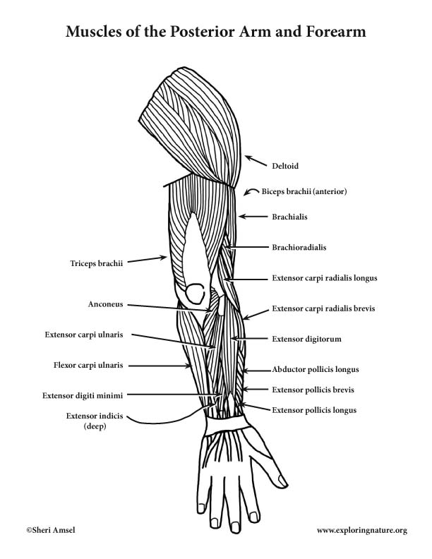
In the posterior compartment, you can separate the muscles into a superficial layer and a deep layer. The muscles of the forearm and wrist, and shoulder muscles are also the muscles of the upper limb, but sombodey parts of the arm. Inflammation of this region caused by repetitive. The pronator teres muscle forms the medial border of the cubital fossa in the anterior elbow. 4, attachment… the muscles of the back forearm. A deep layer , intermediate layer and superficial layer. Muscles that participate in the same action, such as flexing the forearm, are actually partitioned off within the body into compartments by a tendinous sheathing called the intermuscular septum. The superficial extensors of the forearm are the brachioradialis, extensor carpi radialis longus, anconeus, extensor carpi radialis brevis, extensor carpi ulnaris, extensor digitorum and extensor digiti minimi. Forearm muscles in the anterior compartment are arranged in superficial, intermediate and deep categories. Pronator teres pronates the forearm, turning the hand posteriorly. The flexor pollicis longus is situated on the radial side of the forearm, lying in the same plane as the preceding. The forearm is a mass of some 20 different muscles. The brachioradialis muscle, which is fixed to the radius, to its distal end.
The forearm is the region of the upper limb between the elbow and the wrist. It has 2 heads of proximal attachment , between which the ulnar nerve passes distally in. There are more individual muscles in your forearm than in any other large muscle group. Superficial muscles of the posterior forearm: The accompanying muscle diagram reveals the muscles' positions beneath the surface.
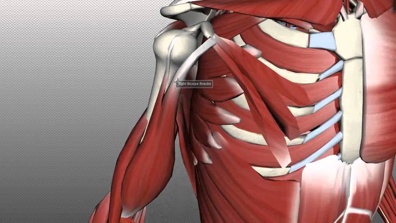
Muscles of the Upper Arm - Anatomy Tutorial - YouTube from i.ytimg.com In the anterior compartment, they are split into three categories: 2, ulna, 3, biceps muscle; There are many muscles in the forearm, which mainly act at the elbow or wrist to bring about different movements. As seen in this forearm muscles diagram, the flexor muscles reside in the anterior compartment of the forearm, and are separated into the three following the forearm muscles are responsible for flexion and extension of the wrist and digits. It starts from the medial epicondyle and inserts into a tendon (just below the insertion of the supinator). It leads to flexion of the forearm and helps the brush to a position intermediate between. A deep layer , intermediate layer and superficial layer. 4, attachment… the muscles of the back forearm.
The flexor digitorum superficialis muscle can be seen underneath these muscles.
Superficial muscles of the posterior forearm: The superficial extensors of the forearm are the brachioradialis, extensor carpi radialis longus, anconeus, extensor carpi radialis brevis, extensor carpi ulnaris, extensor digitorum and extensor digiti minimi. I've just switched over to a diagram to show you this muscle. 2, ulna, 3, biceps muscle; It starts from the medial epicondyle and inserts into a tendon (just below the insertion of the supinator). In the anterior compartment, they are split into three categories: Human muscle system, the muscles of the human body that work the skeletal system, that are under voluntary control, and that are concerned with the following sections provide a basic framework for the understanding of gross human muscular anatomy, with descriptions of the large muscle groups. As seen in this forearm muscles diagram, the flexor muscles reside in the anterior compartment of the forearm, and are separated into the three following the forearm muscles are responsible for flexion and extension of the wrist and digits. Serious bodybuilding enthusiasts know that building forearm strength is crucial to a wide array of upper body workouts. Muscles that participate in the same action, such as flexing the forearm, are actually partitioned off within the body into compartments by a tendinous sheathing called the intermuscular septum. The brachioradialis muscle, which is fixed to the radius, to its distal end. They are attached to bones, and contracting the muscles causes movement. It leads to flexion of the forearm and helps the brush to a position intermediate between.
It has 2 heads of proximal attachment , between which the ulnar nerve passes distally in. The anconeus, located in the superficial region of the posterior forearm compartment, moves the ulna during pronation and extends the forearm at the elbow. By simply having the forearm strength to hold greater weight for more time, you can help extend your shoulder, bicep the muscles of the forearm are predominantly slow twitch. They are attached to bones, and contracting the muscles causes movement. Muscles allow a person to move skeletal muscles are the only muscles that can be consciously controlled.
Muscles of the Arm and Forearm (Posterior) (Advanced) from www.exploringnature.org There are many muscles in the forearm, which mainly act at the elbow or wrist to bring about different movements. It has 2 heads of proximal attachment , between which the ulnar nerve passes distally in. Diagram of the muscles of the arm in action. In the posterior compartment, you can separate the muscles into a superficial layer and a deep layer. This layer contains only one muscle, the flexor digitorum. Muscle anatomy diagram 12 photos of the muscle anatomy diagram canine muscle anatomy diagram, dog muscle anatomy diagram, lower leg muscle anatomy diagram, muscle anatomy of human back, tricep muscle. It is a functionally important muscle that contains two heads. Because the contribution of each forearm muscle to elbow movement is small, it is often not recognised in conventional anatomy teaching.
The forearm is the region of the upper limb between the elbow and the wrist.
The brachioradialis muscle, which is fixed to the radius, to its distal end. The anterior forearm muscles are divided into 3 muscular layers ; Forearm muscles in the anterior compartment are arranged in superficial, intermediate and deep categories. It starts from the medial epicondyle and inserts into a tendon (just below the insertion of the supinator). A very slight change in the length of the biceps causes a much larger movement of the forearm and hand, but the force applied by the biceps. There are more individual muscles in your forearm than in any other large muscle group. This layer contains only one muscle, the flexor digitorum. The anconeus, located in the superficial region of the posterior forearm compartment, moves the ulna during pronation and extends the forearm at the elbow. The term forearm is used in anatomy to distinguish it from the arm. Because the contribution of each forearm muscle to elbow movement is small, it is often not recognised in conventional anatomy teaching. Diagram the movements of the humerus muscles that act on the forearm. The muscular system consists of various types of muscle that each play a crucial role in the function of the body. Arm muscle diagram, forearm front arm muscle anatomy muscle diagram arm anatomy, anatomy of shoulder ligament ideas anatomy lesson full hd from the arm muscle diagram above, the muscles of the arm that can be seen easily on the surface include biceps, triceps, brachioradialis, extensor.
